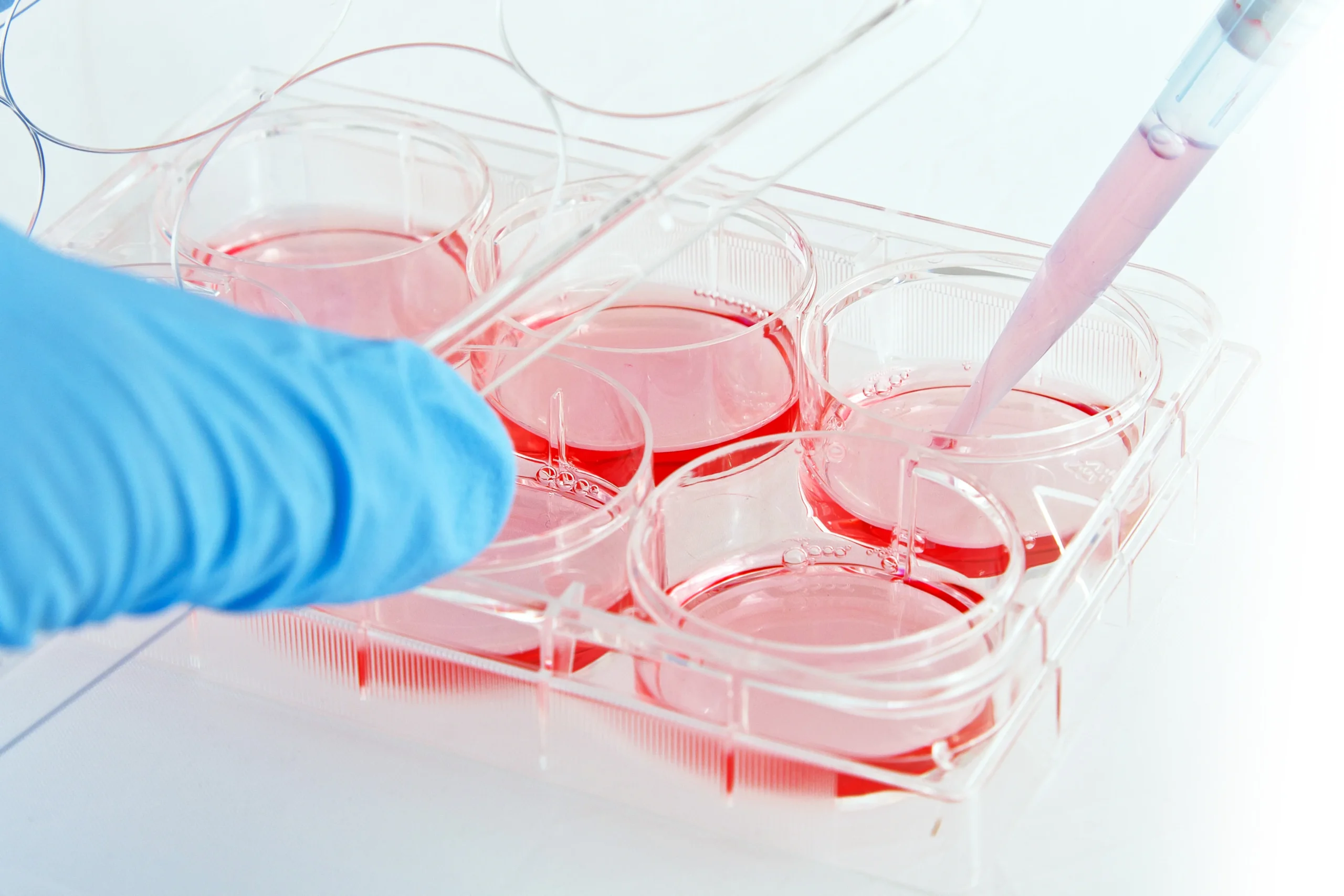
Hepatitis E virus (HEV) is challenging to cultivate in vitro. Replication is often weak and highly strain dependent. Also, viruses released from cells in culture are cloaked in cell-derived envelopes that reduces infection efficiency. Hepatitis E virological research is often conducted using infectious clone derived virus. Clones such as Kernow-C1 have enabled an in-depth examination of HEV that would not have been possible with native virus particles. However, the search for an efficient method for culturing and passaging native HEV from clinical samples is ongoing. In recent times, several groups have attempted to improve HEV yields via modifications in maintenance media such as amphotericin B, MgCl2 (Schemmerer et al, Viruses, 2019), DMSO (Capelli et al, Viruses, 2020) and progesterone (Sooryanarain et al, mBio, 2021). These important works found that HEV growth in vitro was indeed boosted with these modifications, but studies differed in terms of virus strains/ infectious clones, basal media and cell lines used. The team of Dr. Siddharth Sridhar from the University of Hong Kong set out to examine whether these findings were widely generalizable with other HEV strains and cell lines.
In this replication study, the authors examined the impact of the above mentioned cell culture modifications on growth of infected patient stool-derived Paslahepevirus balayani genoype 4 (HEV-A4) and Rocahepevirus ratti genotype 1 (HEV-C1) in PLC/PRF/5 cells. The addition of amphotericin B and MgCl2 clearly boosted yields of HEV-A4 and infected cells could be maintained continuously for 18 months with steady virus production and demonstrable viral protein in supernatant and cell lysates. Virus could also be passaged indicating release of viable virus from cells. However, growth of HEV-C1 was quite poor despite this modification. Yields of HEV-C1 were slightly improved with the addition of 1% DMSO to media already containing amphotericin B and MgCl2. However, the addition of progesterone did not have much additive effect on HEV-A4 or HEV-C1 yields, perhaps indicating a genotype specific effect of this hormone on HEV growth. They also examined the effectiveness of various protocols to remove viral envelope from cell culture derived particles. They found that an NP-40 based protocol was best at removing viral envelope without excessive loss of viable virus.
This study highlights the importance of independent verification using multiple virus strains and liver cell lines to check the generalizability of cell culture modifications for growing HEV in vitro. The authors look forward to increasing use of native patient-derived viruses in HEV research. Recently developed cell culture modifications are a crucial advance in this field.
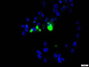
Figure legend: HEV-A infected PLC/PRF/5 cells showing HEV ORF2 protein in cytoplasm. Nuclei stained with DAPI.
Link to study published on 9 June 2022 in Viruses: Independent Evaluation of Cell Culture Systems for Hepatitis E Virus