The hepatitis E virus (HEV) capsid protein ORF2 is best known for its essential role in virion assembly. However, a new study by Jordan & Hu et al., led by Dr. Viet Loan Dao Thi, uncovers an unexpected second function of ORF2: the formation of intracellular amyloid-like fibrils that suppress antiviral immunity and promote viral replication.
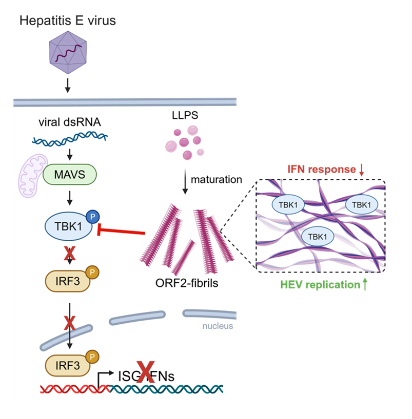
Using a combination of cell biology, biochemistry, confocal microscopy, and correlative light and electron microscopy, the authors discovered that ORF2 forms unconventional filamentous structures in multiple infected cell types and across diverse HEV genotypes. They further demonstrated that the intrinsically disordered N-terminal region of ORF2 drives liquid–liquid phase separation (LLPS), leading to the formation of dynamic biomolecular condensates. Likely facilitated by additional aggregation-prone domains within ORF2, these condensates subsequently mature into filaments that, as revealed by correlative light and electron microscopy, form highly ordered fibrillar assemblies with ultrastructural features reminiscent of amyloid fibrils.
Importantly, the authors show that deletion of the N-terminal intrinsically disordered region impairs HEV replication specifically in immunocompetent cells, indicating that ORF2 fibril formation contributes to viral fitness by antagonizing host antiviral defenses.Mechanistically, the study identifies the innate immune kinase TBK1 as a key target of ORF2 fibrils. While double-stranded RNA sensing remains intact, ORF2 fibrils interact with and sequester TBK1, thereby preventing efficient downstream phosphorylation of IRF3 and suppressing antiviral gene expression. These findings provide a mechanistic explanation for previous observations that ORF2 counteracts cell-intrinsic antiviral responses during HEV infection.
More broadly, this work expands the growing repertoire of viruses that exploit biomolecular condensates and phase transitions during infection. Similar higher-order assemblies have been described for proteins from several unrelated viruses, yet their biological functions often remain incompletely understood. In the context of HEV infection, ORF2 fibrils appear to function as specialized intracellular platforms that spatially reorganize antiviral signaling pathways, thereby creating a more permissive environment for viral replication.
The study also opens several exciting avenues for future research. High-resolution structural studies will be required to define the molecular architecture of ORF2 fibrils and to understand how fibril formation relates to the canonical capsid-forming function of ORF2. In addition, it will be important to determine whether ORF2 fibrils, analogous to other viral protein-derived amyloids, contribute directly to HEV-associated pathogenesis and disease progression.
Read the full article: Cell Rep. 2026 June 23;45(6):117513. DOI: 10.1016/j.celrep.2026.117513