At least 20 million hepatitis E virus (HEV) infections occur annually. Hepatitis E is generally acute and self-limiting. However, chronic hepatitis E has gained more attention worldwide and has been recognized as an emerging significant clinical challenge not only confined to immunocompromised patients but also to immunocompetent individuals. The lack of a desirable small animal model for chronic HEV infection hinders our understanding of the pathogenesis and the development of therapeutics measures.
A recent study led by the group of Prof. Youchun Wang and Prof. Chunnan Liang from National Institutes for Food and Drug Control, China, investigated chronic HEV infection in immunocompetent rabbits. They characterized the course of chronic HEV infection in immunocompetent rabbits. Fecal virus shedding in rabbits is the most sensitive and stable marker of HEV infection and can appear as early as one week post-infection (wpi). All rabbits that developed chronic hepatitis E had persistent fecal virus shedding till the end of the study at 26 wpi. Serum viral RNA of the infected rabbits persisted for a much shorter period of time when compared to fecal virus shedding.
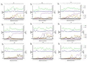
They also investigated the kinetics of HEV-Ag in the serum and fecal samples of the rabbits as the long-term kinetics of HEV Ag detection during HEV infection is not known. It is interesting that most of the rabbits that developed chronic hepatitis E present with persistent antigenemia at the early phase of the infection. However, those rabbits that spontaneously cleared the infection within 8 wpi showed almost no HEV antigenemia or fecal antigen shedding during the whole experiment. A dramatic increase of HEV-Ag level in serum and feces of most of the chronically infected rabbits was observed after 13 wpi, which suggested accumulation of a secreted form HEV capsid proteins and active HEV replication at the chronic phase of infection.
The dynamics of liver histopathology was studied in the present immunocompetent rabbit model. At the acute phase, inflammation and lymphocyte infiltration is the major observation with spotty necrosis. At the chronic phase, liver tissues of rabbits showed features of chronic viral hepatitis. These findings are similar to chronic hepatitis E patients. Characterizations of chronic HEV infection in immunocompetent settings could be recapitulated in rabbits, which can serve as a valuable tool for future studies on pathogenesis. The chronic lesions in the liver were also supported by serum biochemical analysis of liver enzymes. The level of AST showed persistent mild-to-moderate elevation in most of the chronically infected rabbits during the whole study period, indicating ongoing liver injury. GGT level was significant higher in rabbits with chronic hepatitis E than that of the recovery rabbits at 26 wpi. This study provided a promising tool for future study of the pathogenesis and underlying mechanism for why chronic HEV can also occur in immunocompetent individuals.

Figure . Dynamic of HEV markers and liver function tests in rabbits with chronic HEV infection. A5, B4, C1, D2, E2, F3, G2, H4 and I3 are the number of each representative rabbit. Detection of HEV RNA, HEV antigen, liver function tests, and anti-HEV antibody detection in nine representative chronically infected rabbits, one rabbit per group. Serum and fecal samples were collected weekly for immediate analysis. “-” for negative and “+” for positive of HEV RNA detection by RT-qPCR. Ab, antibody; Ag, antigen; ALT, alanine aminotransferase; AST, aspartate aminotransferase; B, blood; F, feces; HEV, hepatitis E virus; WPI, weeks post-inoculation.
This study was published in Viruses in June 2022. Read the full article https://www.mdpi.com/1999-4915/14/6/1252

